![]()
In-Vivo Imaging
|
Endoscopic imaging for minimal access surgery has many limitations
that include: 2D and narrow angle imaging, limited workspace of
the endoscope caused by the fulcrum effect of the body wall, and
the presence of the endoscope in the incision that prevents use
of the incision for other instrumentation. We have designed a novel
stereoscopic 3D imaging device with 5 DOF and remote control that
can be inserted and attached in the body cavity. The device, contained
within a 11/16" tube, includes two miniature cameras and five
small motors that position the cameras to provide a stereoscopic
view of the surgical site. When inserted the cameras are retracted
and protected by an outer shell. After the device is fixed within
the abdominal cavity, a motor rotates an inner shell to expose the
cameras. Once exposed, the cameras can tilt in tandem, translate
independently along the axis of the tube, and independently pan.
The software controls the cameras to create new views for the surgeon,
to move along the adjustable baseline, to verge for stereoscopic
viewing, and to potentially track moving organs. We have completed
a proof of concept design, which includes CAD models and animations
of the device, and we are currently building a physical prototype.
Once the prototype is completed, we will begin testing it in a surgical
mock-up, followed by animal and clinical trials.
[Patent pending]
Andrew Miller, Peter Allen, and Dennis Fowler. “In-vivo stereoscopic imaging system with 5 degrees-of-freedom for minimal access surgery.” In Medicine Meets Virtual Reality 12, pp. 234-240, January, 2004. |
Powerpoint presentation from the 12th Medicine Meets
Virtual Reality Conference, 2004. |
|
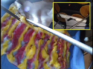
camera4.avi - Early simulation model of the camera system that shows the degrees of freedom of the device.
take2.avi - Shows motion of two of the DOF of the first camera module. take2divx.avi - Higher quality version of the same move, but requires dvix codec to view.
demo4.mpg - Shows picture in picture view of the image from the device and the motions it makes. An experiment is performed in a laparoscopic training box. |